Espondilolistesis y espondilólisis, caso clínico radiológico
Palabras clave:
espondilolistesis; espondilólisis; dolor de la región lumbar; articulación cigapofiaria; rayos X; imagen por resonancia magnética; tomografía computarizada multidetector.Resumen
Introducción: con frecuencia son atendidos cientos de pacientes en las clínicas de dolor afectados con lumbago, originado por disimiles causas.
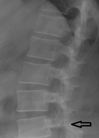
Caso clínico: paciente joven de 37 años con dolor lumbar causado por defecto degenerativo en la faceta articular conocido por espondilolistesis, diagnosticado por los protocolos de estudios radiológicos con uso de estudio convencional de rayos X de columna lumbar en proyección lateral. Muestra ligeros cambios de osteoartritis, desplazamiento anterior incipiente de L4 y solución de continuidad a nivel de la faceta articular inferior L4, un estudio de resonancia practicado nos muestra adicionalmente que la faceta esta engrosada, compromete el diámetro del foramen correspondiente con compresión del ganglio raquídeo L5 del lado izquierdo, y para evaluar y confirmar la afección se realizó exploración con tomografía computarizada de columna lumbar con reconstrucción sagital en un tomógrafo multidetector. Se evidencia claramente solución de continuidad a nivel de la cortical ósea de la faceta inferior izquierda L4.
Conclusión: la espondilolistesis y espondilólisis son bastante frecuentes y causan dolor lumbar bajo. El estudio convencional de rayos X de columna lumbar resulta útil en el diagnóstico, pronóstico y evaluación de espondilolistesis y espondilólisis. La resonancia magnética nuclear y la TAC brindan información adicional sobre las estructuras afectadas, y es más útil en paciente con ciática y grado bajo de espondilólisis.
Descargas
Citas
1. Guille O, Challier V, Parent H, Cavagna R, Poignard A, Faline A, et al. Degenerative lumbar spondylolisthesis. Cohort of 670 patients, and proposed of a new classification. Orthop Traumatol Sur Res. 2014; 100:311-5.
2. Moreaus PE, Ferrero E, Rionallon G, Lenoir T, Guigui P. Radiologic adjacent segment degeneration 2 years after lumbar fusion for degenerative spondylolisthesis. Orthop Traumatol Surg Res. 2016; 102:759-63.
3. Moreau PE, Flouzart Lacharette CH, Lebhar J, Mirouse G, Puignard A. Particularities of anterior fusion in L4-L5 isthmic spondylolisthesis. Orthop Traumatol Surg Res. 2016; 102:755-8.
4. Zhang S, Ye C, Lai Q, Lui X, Nie T, et al. Double-level lumbar spondylolysis and spondylolisthesis: A retrospective study. J Orthop Surg Res. 2018; 13:55.
5. Kang EK, Park H, Kim SH, Baek S. Clinical Usefulness of X-Ray Findings for Non-specific Low Back Pain in Korean Farmers: FARM study. Ann Rehabil Med. 2017;41(5):808-15.
6. Valls Pérez O, Parrilla Delgado ME, Valls Figueroa CT. Evaluación imaginológica en pacientes con sospecha de lumbalgia aguda. En: Imaginología de urgencia. Valor de los algoritmos diagnósticos. TII. La Habana: Editorial Ciencias Médicas;2012. p.684-92.
7. Bravo Acosta. Afecciones de la columna dorsolumbar. En Diagnóstico y rehabilitación en enfermedades ortopédicas. La Habana: Editorial Ciencias Médicas; 2016.p. 280-97.
8. Perera RS, Dissanayake PH, Senarath U, Wijayaratne LS, KarunanayakeAL, Dissanayake VH. Association between disc space narrowing, anterior osteophytes and disability in chronic mechanical low pain: a cross sectional study. BMC Musculoesk Dis. 2017; 18:193.
9. Capote Cabrera A, López Pérez YM. Rayos X. En Medios diagnósticos imaginológicos en rehabilitación. La Habana: Editorial Ciencias Médicas;2011. p. 1-120.
10. Capote Cabrera A, López Pérez YM. Tomografía axial computarizada. En Medios diagnósticos imaginológicos en rehabilitación. La Habana: Editorial Ciencias Médicas;2011. p. 162-182.
11. Capote Cabrera A, López Pérez YM. Resonancia magnética nuclear. En Medios diagnósticos imaginológicos en rehabilitación. La Habana: Editorial Ciencias Médicas;2011. p. 183-215.
12. Petracchi MG, Camino Willhuber G, González Viescas JM, de Cicco FL, Gruenbrg M, Sola C. Osteosíntesis directa para tratar la espondilolistesis traumática del axis. Reporte de caso y revisión de literatura. Rev Assoc Argent Ortop Traumatol. 2016; (Suppl):23-7.
13. Guyot JP, Zaragoza E, Lloyd R, Furmento R, Gelosi F. Espondilolistesis traumática lumbosacra. Reporte de cuatro caos y revisión de la bibliografía. Rev Assoc Argent Ortop Traumatol. 2017; 82(3):249-52.
14. Yapeng JS, Hui W, Dalong Y, Nan Z, Sidong Y, Wei Z, et al. Characterization of radiographic fetures of consecutive lumbar spondylolisthesis. Medicine. 2016; 95(46): e5323.
15. Guiroy A, De Boris G, Jalón P, Gagliardo M, Reviriego J, Rositto G. Espondilolistesis traumática L5-S1: presentación de 3 casos. Rev Argent Neuroc [Internet].2016 [citado 22 Feb 2018]; 30(1). Disponible en: http://aanc.org.ar/ranc/items/show/146.
16. Tao BW, Hui W, Huan L, Lei M, Feng-Yu L, Wen-Yuan D. Sagittal spinopelvic parameters in 2-level lumbar degenerative spondylolisthesis: A retrospective study. Medicine. 2016;95(10): e5417.
Descargas
Publicado
Cómo citar
Número
Sección
Licencia
Avisos de derechos de autor propuestos por Creative Commons
1. Política propuesta para revistas que ofrecen acceso abierto
Aquellos autores/as que tengan publicaciones con esta revista, aceptan los términos siguientes:- Los autores/as conservarán sus derechos de autor y garantizarán a la revista el derecho de primera publicación de su obra, el cuál estará simultáneamente sujeto a la Licencia de reconocimiento de Creative Commons que permite a terceros compartir la obra siempre que se indique su autor y su primera publicación esta revista.
- Los autores/as podrán adoptar otros acuerdos de licencia no exclusiva de distribución de la versión de la obra publicada (p. ej.: depositarla en un archivo telemático institucional o publicarla en un volumen monográfico) siempre que se indique la publicación inicial en esta revista.
- Se permite y recomienda a los autores/as difundir su obra a través de Internet (p. ej.: en archivos telemáticos institucionales o en su página web) antes y durante el proceso de envío, lo cual puede producir intercambios interesantes y aumentar las citas de la obra publicada. (Véase El efecto del acceso abierto).