Congenital myelomeningocele. Case presentation
Keywords:
Myelomeningocele; Ventricular dilation and hydrocephalus.Abstract
Introduction: myelomeningocele is a congenital anomaly of the neural tube in which the bones of the spine are not fully formed and the spinal canal is incomplete, which allows the spinal cord and meninges to protrude through the child's back.
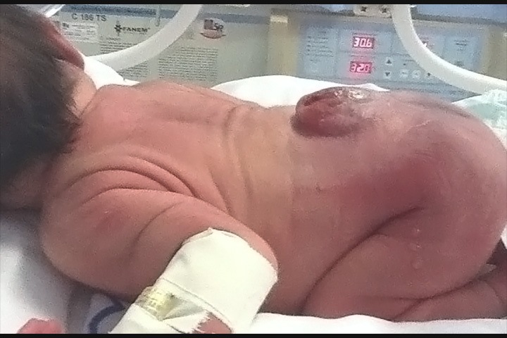
Case presentation: newborn male, product of a dystocic delivery (cesarean section by pelvic plus prenatal diagnosis of congenital malformation), clinically at term (EG 38 weeks by Parkin test), with good birth weight (3 600 gr) , Apgar 9-9, Nonreactive Serology, Group and Factor B positive, with cord and normal placenta, a clear amniotic fluid and without RPM. On physical examination, he presents a full sac that protrudes in the lumbosacral region accompanied by total paralysis of both lower limbs. No signs of respiratory distress, pink, well perfused, crying loudly and preserved vitality.
Discussion: 22-day-old newborn who is taken to the operating room to perform a ventricle-peritoneal shunt with the diagnosis of active hydrocephalus. A sample of the cerebrospinal fluid was previously taken for negative studies. It was decided to associate it with treatment with acetozolamide or glaumox and up to now it has maintained a satisfactory evolution.
Conclusions: the newborn presents various symptoms and signs common to a malformation of the central nervous system (Congenital Myelomeningocele); In this particular case it is striking that, similar to what is reported in the literature on the subject, the neonate presented ventricular dilation and associated Hydrocephalus.
Downloads
References
1. Calderón Yanes M, Mesa Suarez M, Marrero Escobedo D. Defecto del tubo neural. Presentación de caso. Rev Cubana Obstetr Ginecol 2017; 43(1): 1-7.
2. Bidondo Maria P, Liascovich R, Babero P, Groesman B. Prevalence of neural tube defects and estimation of cases overted in the post-fortification period in Argentina. Arch Arg Pediatr 2015; 113(6): 498-501.
3. BPS. Instituto de Seguridad Social. Guía Clínica. Diagnóstico y Tratamiento Mielomeningocele. [Internet]. Montevideo: Instituo de Seguridad Social; 2015. [Citado 23/2/2019]. Disponible en: https://www.bps.gub.uy/bps/file/13117/1/guia_clinica_mielomeningocele.pdf
4. Sani S Maldenhauer JS, Sprinner SS, Rendon N, Khaelek N et al. Chorioamniotic membrane separation preterm premature rupture of membranes complicating in utero myelomeningocele repair. Am J Obstet Gynecol 2016; 214(5): 647.e1-7.
5. González Pérez F, Águila Hernández Y, Ibáñez Palacio V, Jiménez Hernández V. Diagnóstico de myelomeningocele en un feto mediante resonancia magnética de bajo campo. Presentación de un caso. Medisur 2018; 16(1): 85-89.
6. Belfort MA, Whitehead WE, Shamshirsaz AA, Ruano R, et al. Fetoscopic repair of Meningomyelocele. Obstet Gynecol 2015; 126(4): 881-4.
7. Volpe JJ. Human Brain Development. In: Volpe JJ. ed. Neurology of the Newborn. 3rd ed. Philadelphia: Saunders; 2015: p. 3.
8. Martinez Sabater A. Arnold-Chiari malformation: loss smile. Index Enferm [Internet]. 2014 [citado 29/9/2019]; 23(4). Disponible en: http://scielo.isciii.es/scielo.php?script=sci_arttext&pid=S1132-12962014000300013
9. Pedreira DA, Nelci Zanon L, Nishi Kuni K, Moneire de Sara Acaceo GL et al. Endoscopic surgery for the antenatal tratement of myelomeningocele. The CECAM trial. Am J Obstet Gynecol 2016; 214(1): 111.e1-111.e11.
10. Hernández Ruiz I, Soler Cano A. Malformación de Arnold Chiari tipo I: presentación de un caso. Rev. Méd. Electrón. [Internet]. 2010 [citado 12/2/2019]; 32(5). Disponible en: http://www.revmedicaelectronica.sld.cu/index.php/rme/article/view/757/html
11. Amado Vázquez ME, García Ramos R, Avellaneda Fernández A, García Ribes M, Barrón Fernández J, Gómez Triguero C, et al. Malformaciones de la unión cráneo-cervical (Chiari tipo I y siringomielia). Documento de consenso. [Internet]. Madrid: Editorial Médica AWWE; 2009. [citado 12/6/2019]. Disponible en: http://www.sen.es/pdf/2010/Consenso_Chiari_2010.pdf
12. Gregorian M. Defectos del Tubo Neural: En: Lane H. Manual de Pediatría. Madrid: Mosby-Doyma; 2015: p. 398.
13. García CR. Defectos del Tubo Neural: una responsabilidad compartida. En: PAC Neonatología. Programa de Actualización Continua en Neonatología. [Internet]. México: Federación Nacional de Neonatología de México; 2017. [citado5/9/2019]. Disponible en: https://www.anmm.org.mx/publicaciones/PAC/PAC_Neonato_4_L2_edited.pdf
14. Delgado GC. Malformaciones Congénitas. En: PAC Neonatología. Programa de Actualización Continua en Neonatología. [Internet]. México: Federación Nacional de Neonatología de México; 2017. [citado5/9/2019]. Disponible en: https://www.anmm.org.mx/publicaciones/PAC/PAC_Neonato_4_L2_edited.pdf
Downloads
Published
How to Cite
Issue
Section
License
Avisos de derechos de autor propuestos por Creative Commons
1. Política propuesta para revistas que ofrecen acceso abierto
Aquellos autores/as que tengan publicaciones con esta revista, aceptan los términos siguientes:- Los autores/as conservarán sus derechos de autor y garantizarán a la revista el derecho de primera publicación de su obra, el cuál estará simultáneamente sujeto a la Licencia de reconocimiento de Creative Commons que permite a terceros compartir la obra siempre que se indique su autor y su primera publicación esta revista.
- Los autores/as podrán adoptar otros acuerdos de licencia no exclusiva de distribución de la versión de la obra publicada (p. ej.: depositarla en un archivo telemático institucional o publicarla en un volumen monográfico) siempre que se indique la publicación inicial en esta revista.
- Se permite y recomienda a los autores/as difundir su obra a través de Internet (p. ej.: en archivos telemáticos institucionales o en su página web) antes y durante el proceso de envío, lo cual puede producir intercambios interesantes y aumentar las citas de la obra publicada. (Véase El efecto del acceso abierto).