Valor predictivo de la tomografía en el diagnóstico de los astrocitomas cerebrales. Hospital Carlos Manuel de Céspedes. 2017-2018
Palabras clave:
Tomografía; Imagen por resonancia magnética; Neoplasias encefálicas.Resumen
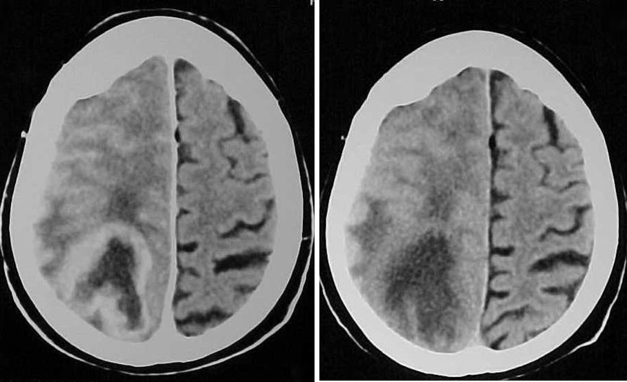
Los astrocitomas son tumores cerebrales con una elevada frecuencia de aparición. Se realizó un estudio no experimental con componente descriptivo y analítico, con el objetivo de determinar el valor predictivo de las variables tomográficas en el diagnóstico de los astrocitomas cerebrales en 52 pacientes con diagnóstico tomográfico de neoplasia cerebral confirmado por Tomografía Axial Computarizada (TAC) de cráneo atendidos en el Hospital Carlos Manuel de Céspedes de Bayamo en el período comprendido entre enero del 2017 hasta diciembre del 2018. En el presente estudio encontramos que la región más frecuentemente afectada por astrocitomas fue la frontoparietal seguida de la región frontal y temporal. El estudio tomográfico diagnosticó 23 pacientes con el diagnóstico de astrocitomas y 29 como no astrocitomas, se encontró una alta sensibilidad y especificidad, así como valores predictivos positivos y negativos altos. En cuanto a las variables tomográficas evaluadas, el realce de contraste escaso y la hipodensidad tuvieron alta sensibilidad y especificidad, así como sus valores predictivos de lo que resulta que son características útiles en el diagnóstico. El índice de validez calculado tanto en la tomografía como en las variables estudiadas en sentido general aporta alta veracidad.
Descargas
Citas
1. Den Hollander M, Bensch F, Glaudemans A, Oude Munnink T, Enting R, Walenkamp A, et al. TGF-β Antibody Uptake in Recurrent High-Grade Glioma Imaged with 89Zr-Fresolimumab PET. J Nuc Med 2015; 56(9): 1310-1314.
2. Fu X, Sun Z, Chang Y. Expression and clinical significance of P53, O6-methylguanine-dna methyltransferase and epidermal growth factor receptor in glioma. J Biol Regul Homeost Agents 2015; 29(4): 853-858.
3. Cala Irén M, Pons Porrata LM, Domínguez Piorno R, Salomón López J. Caracterización clinicohistopatológica, tomográfica y por resonancia magnética de pacientes menores de 15 años con tumores cerebrales. MEDISAN 2017; 21(7): 797-804.
4. Zhu X, Mou K, Xu Q, Tang J, Huang G, Lv S, et al. Microarray analysis of the aberrant microRNA expression pattern in gliomas of different grades. Oncology Reports 2015; 34(1): 318-324.
5. Castañeda CA, Casavilca S, Orrego E, García Corrochano P, Deza P, Heinike H, et al. Glioblastoma: Análisis molecular y sus implicancias clínicas. Rev Peru Med Exp Salud Pública 2015; 32(2): 316-325.
6. Martín Martínez Y, García Amelo IM, Hernández Viel Valia, Miranda Mustelier N, Domínguez Piorno R. Valor de la tomografía axial computarizada para el diagnóstico de tumores craneales supratentoriales MEDISAN 2013; 17(2): 237-305.
7. Sierra Benítez EM, León Pérez MQ, Laud Rodríguez L, Carrillo Comas AL, Pérez Ortiz L, Rodríguez Ramos E. Gliomas malignos: biología molecular y detalles oncogenéticos. Rev Médica Electrónica 2018; 40(4): 1100-1111.
8. Hathout L, Ellingson B, Cloughesy T, Pope W. Patient-specific characterization of the invasiveness and proliferation of low-grade gliomas using serial MR imaging and a mathematical model of tumor growth. Oncology Reports 2015; 33(6): 2883-2888.
9. Kros J, Huizer K, Hernández-Laín A, Marucci G, Michotte A, Gorlia T, et al. Evidence-Based Diagnostic Algorithm for Glioma: Analysis of the Results of Pathology Panel Review and Molecular Parameters of EORTC 26951 and 26882 Trials. J Clin Oncol 2015; 33(17): 1943-1950.
10. Posti J, Bori M, Kauko T, Sankinen M, Nordberg J, Sipilä J, et al. Presenting symptoms of glioma in adults. Acta Neurol Scand 2015; 131(2): 88-93.
11. Chaumeil M, Lupo J, Ronen S. Magnetic Resonance (MR) Metabolic Imaging in Glioma. Brain Pathol 2015; 25(6): 769-780.
12. Wang J, Ma Y, Cooper MK. “Cancer stem cells in glioma: Challenges and opportunities”. Transl Cancer Res 2013; 2(5): 429-441.
13. Mequins LC, Adry RA, Silva Júnior SC, Pereira CU, Oliveira JG, Morais DF, et al. Gross-total resection of temporal low grade gliomas is a critically important factor in achieving seizure-freedom. Arq NeuroPsiquiatr 2015; 73(11): 924-928.
14. Arbizu J, Domínguez PD, Diez R, Vigil C, García R, Zubieta JL, et al. Neuroimagen de los tumores cerebrales. Rev Española Med Nucl 2011; 30(1): 47-65.
15. Saldívar-Rodea CA, Guerrero-Avendaño GM, Benítez-Barradas MI, Reyes-Caldelas MA. Utilidad de la resonancia magnética en el diagnóstico y clasificación de los tumores astrocíticos. Anales de Radiología México 2016; 15(4): 279-293.
Descargas
Publicado
Cómo citar
Número
Sección
Licencia
Avisos de derechos de autor propuestos por Creative Commons
1. Política propuesta para revistas que ofrecen acceso abierto
Aquellos autores/as que tengan publicaciones con esta revista, aceptan los términos siguientes:- Los autores/as conservarán sus derechos de autor y garantizarán a la revista el derecho de primera publicación de su obra, el cuál estará simultáneamente sujeto a la Licencia de reconocimiento de Creative Commons que permite a terceros compartir la obra siempre que se indique su autor y su primera publicación esta revista.
- Los autores/as podrán adoptar otros acuerdos de licencia no exclusiva de distribución de la versión de la obra publicada (p. ej.: depositarla en un archivo telemático institucional o publicarla en un volumen monográfico) siempre que se indique la publicación inicial en esta revista.
- Se permite y recomienda a los autores/as difundir su obra a través de Internet (p. ej.: en archivos telemáticos institucionales o en su página web) antes y durante el proceso de envío, lo cual puede producir intercambios interesantes y aumentar las citas de la obra publicada. (Véase El efecto del acceso abierto).